Waterloo Chiropractor, Waterloo Physiotherapist, and Massage Therapist (RMT)

Category

Injuries

By: SAYAKA TIESSEN, HONS. BKIN, MSCPT

Book here

Ankle sprains, AKA rolled ankles, have got to be one of the most common injuries out there. They can happen in the most innocuous situations, like stepping on an uneven surface, or during sports that require cutting and landing. The words “walk it off” are synonymous with ankle sprains. Get up. It’s nothing serious. Back to business.

I get the sense that most people that sustain an ankle sprain do not seek any guidance from a physiotherapist. It might swell up nicely for a day or two, but then you limp around and manage to get on with your life. What you might not appreciate though, is that ankle sprains have a high recurrence rate due to the residual effects of the initial injury. Things like ligament laxity and damage to the neural and musculotendinous tissue around the ankle can inhibit complete recovery. This can present as weakness, poor balance, and slower muscle response to load. You might not notice these deficits when you are walking around, but they do make you susceptible to re-injuring the area. This is why I always advocate for getting any old rolled ankle assessed by a physiotherapist.

I often prescribe a blend of resistance and balance training for my ankle sprain patients. But if you are super crunched for time, is there one that is more efficient than the other?According to a 2021 study, although resistance training and balance training on their own improves strength, hopping (explosiveness and control), and dynamic balance, if you had to pick one, balance training takes the edge. This is likely due to the high training load used in the study (20-30 second holds, 10-20x, 3-5 sets over multiple exercises), and the fact that you need to be strong and coordinated to balance well, and strictly strengthening will not challenge your proprioception (balance-ability). Time to get your Karate Kid on.

Want to know what kind of exercises are appropriate for you? Or have you sprained your ankle before and feel things are not quite right? You can book an appointment with me here!

By: Sayaka Tiessen, Hons. BKin, MSc (PT)

It’s been a while! Life has been insanely busy the past couple months. You might have noticed on my social media feeds that I have moved my physiotherapy practice to join forces with Delanghe Chiropractic & Health/Health & Performance. Pumped for what this opportunity will bring!

So what’s on the agenda today? In honour of a few patients I have seen recently for a variety of running related injuries, we will be addressing the question: do injured runners run differently?

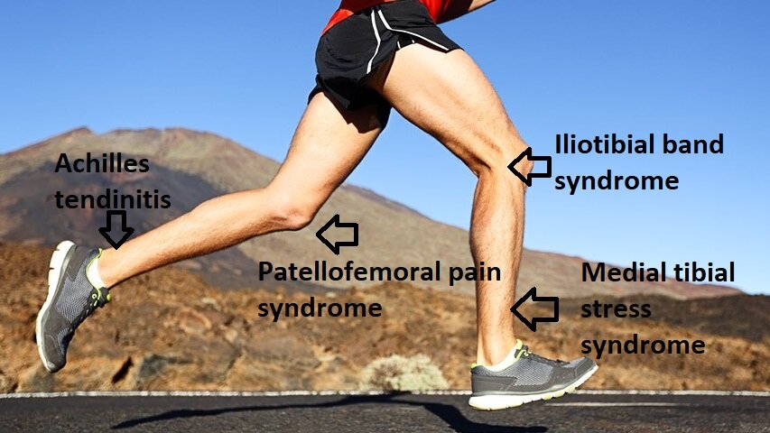
I will be drawing from an article by Christopher Bramah published in 2018 that looks at this very issue. He compared healthy runners (no reported injury in over 18 months) to injured runners to see if there were any run gait characteristics that were predictive of current injury. He specifically looked at the four most commonly cited soft tissue injuries in the running population: Patellofemoral Pain Syndrome, Iliotibial Band Syndrome, Medial Tibial Stress Syndrome, Achilles Tendinitis.

(Let’s just acknowledge the fact that I haven’t used Microsoft Paint in possibly a decade. How is it still a thing?)

You might not have heard of these conditions before, but you have probably experienced them. Here is a quick breakdown of what each of these injuries involve in the simplest of terms:

Patellofemoral Pain Syndrome (PFPS): pain around the patella, aka the knee cap at the front of the knee

Iliotibial Band Syndrome (ITBS): pain at the outside of the knee

Medial Tibial Stress Syndrome (MTSS): AKA Shin Splints (the bane of my existence for many years), pain around the inner part of the bottom 1/3 of the shin.

Achilles Tendinitis (AT): pain at the Achilles tendon.

I have to say, it’s always pretty neat when the findings of a study are similar to what you experience clinically. After analyzing the running biomechanics of the injured and non-injured study participants, Bramah’s team found that the injured runners presented with:

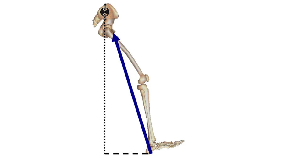
overstride.jpg
A straighter knee and ankle dorsiflexion (toes pulled up to the shin) when landing. This is essentially what we would call overstriding/overreaching while running – your foot is way out in front of your center of mass when you first hit the ground.
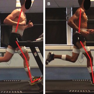
forward lean.jpg
Increased forward trunk lean. This can be caused by low back and gluteus maximus weakness/fatigue. We need to strike a balance of leaning forward too much and sitting back like you’re in a La-Z-Boy. Photo is from the Bramah et al. 2018 article.
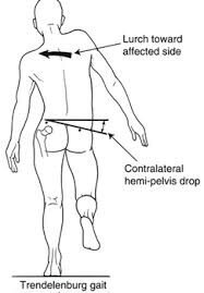
trendelenburg.jpg
A significant drop in the opposite hip to the one they are standing on, also known as a Trendelenburg. This can be a sign of weakness of the hip stabilizing muscles on the leg they are standing on because they cannot keep the pelvis level. Note that the hip drop was found to be the most accurate at predicting if the runner was injured.

These are pretty important to note because they can actually exacerbate your injury by adding stress to the already injured tissues. Talk about adding insult to injury!

I do have to acknowledge that not every runner is the same. There are some runners that are FAST, have a wicked hip drop, and are totally functional. But as a physiotherapist, it is good for me to keep in mind that these running traits are often present with an injured runner.

What are the implications?

If you are a runner dealing with any type of injury, come in and get assessed. The weather is warming up and nothing is more of a bummer than not being able to get out there and enjoy it.

I do not believe that all runners should run the same way. However, I am not opposed to tweaking your run form to help iron out some of these movements patterns. Gait assessments are a great way to get a second set of eyes on your run form and see if there is anything we can adjust with cueing.

On top of running tips, I always give some homework to help you move better. I typically assign a short list of exercises that are targeted to your concerns – all business, no filler.

Don’t let nagging injuries keep you on the couch! Let’s work together to get you back out there! Click here to book now

By: SAYAKA TIESSEN, HONS. BKIN, MSCPT

In both kinesiology and physiotherapy school, we were taught anatomy. The sheer volume of knowledge was overwhelming. Where does each muscle attach? What nerve controls which muscle? It felt like we were learning everything there was to know about the body. That was incredibly naive of me, as research has plowed on and has shown just how complex our movement systems are.

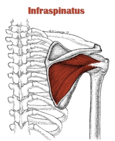
This post will take a closer look at the infraspinatus, one of your four rotator cuff (RC) muscles. Located just under the boney ridge of your shoulder blade, it is commonly injured, especially in overhead athletes (throwers, climbers, etc).

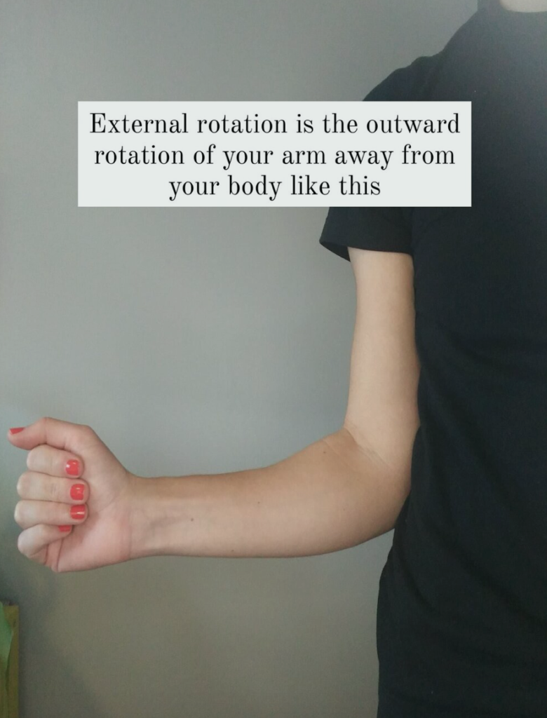
When I learned about the infraspinatus, we were told that it was a muscle that externally rotates, or turns your arm outward (see below for a picture showing that position) and that it is controlled by your suprascapular nerve. But within the last 20 years or so, researchers have found that the infraspinatus has three distinct regions, each innervated by its own mini branch of the suprascapular nerve; the superior, middle, and inferior infraspinatus subregions.

Why does this matter? Well, turns out that the subdivisions serve slightly different purposes, kind of like how your municipality functions within the province. The province of Ontario has an overarching goals, but Waterloo Region will function in a different way than say the GTA. They also will take on more or less burden depending on the task at hand (regional containment of COVID19 being a prime and timely example). This goes for the subregions too – some sections might turn on more or less depending on the degree of arm elevation, your plane of movement, and resistance.


If that’s the case, then is there a way to make rehab more specific by targeting movements that bias one subregion over another?

I was hoping the answer would be yes, but the research isn’t there yet. It seems as though there are still some discrepancies in the research about which subregion does what. Furthermore, the role of the inferior infraspinatus has yet to be determined.

I have summed up 4 key takeaways below in terms of the roles of the subregions:

  1. All three subregions of infraspinatus are more active the higher your arm is in front of you (eg: they will work harder if you are reaching into a high cupboard, and will work less if reaching for something at waist height).
  2. The superior infraspinatus has a shared insertion on the top of the arm bone with your supraspinatus (another RC muscle that will have its own blog post next). It is thought that both muscles contribute to shoulder stabilization, and that redundancy allows for people to have tears and still be strong.
  3. The middle infraspinatus is more of a pure external rotator.
  4. There is minimal evidence for the role of the inferior infraspinatus.

What does this mean for rehab?

  • When you are dealing with a fresh injury, start with exercises where your arm is low and closer to your body, like farmer’s carries. Any load on your arms will turn on your rotator cuff, and this is the least provoking position to be in.
  • You can start with isometrics (exercises where you are not moving your arm through range) if you cannot even move the shoulder without pain.
  • You don’t have to rotate your shoulder to get your infraspinatus. You can just do an arm raise and it will work all three subregions. The higher your arm, the more the infraspinatus will be working.
  • Generally, strengthening external rotation follows the same principle. The movement of rotating your arm outward will be more challenging the further away your upper arm is from your body (overhead vs tucked into your side).

Check out the video below for 4 different exercises that target the infraspinatus, ranging from lower muscle activation to the most, using the principles I outlined above. Some of these exercises are based on articles published by researchers at the Digital Industrial Ergonomics and Shoulder Evaluation Laboratory at the University of Waterloo (local shout out!).

The next write up will target the supraspinatus – yet another notorious RC muscle that also has 2 subregions despite being super slender. As I eluded to above, superior infraspinatus shares an insertion with supraspinatus, and we will talk about why that is so important for shoulder stability and how you could approach rehabilitation for a torn supraspinatus. Stay tuned!

Sayaka is an evidence based physiotherapist in Waterloo. Click here

to learn more about her!

How long until we officially race again?  I would say probably at least a few weeks, wouldn’t you?  While I’m sure many of you miss toeing the line, there are some positives.  For instance, one of the best ways to take advantage of this extended offseason is to work on weaknesses that are normally tough to address.

Constantly acting in A-race mode, followed by tapering, followed by recovering can definitely result in short term spikes in performance. But often the long term, gradual development is sacrificed.

A great way to take a swing at improving your baseline ability to perform is a full strength program. However, it is not as logical to include when in close proximity to an A-race.  If you’re anything like me, you’ve thought of introducing more strength work throughout the pandemic.  If you’re not like me, you’ve actually done it- good for you, you jerk!

The questions that I get from the team on this topic are endless.  I have written about the topic in short before such as here.  However, I thought it was finally time to take a deep dive into strength training for endurance athletes.

CLICK HERE to read the rest in the Run Waterloo Magazine. 

We all get injured.  Muscle strains. Tendonitis. Stress fractures. The list goes on for runners! Not to mention we also have to deal with all the regular life injuries that others have to cope with like headaches, lower back pain and shoulder issues.

Yet, doesn’t it seem that certain people recover faster from injury than others?  Even if two people have exactly the same injury, and do exactly the same things to manage it, the healing time can still be drastically different.

So what separates us?  There is no doubting that physical characteristics, such as age and level of fitness, can impact recovery time.  However, if all things physical are held equal, healing times for the same injury can STILL differ drastically.

Why is this? One possible explanation: the influence of the mind.

CLICK HERE to read the rest in the Run Waterloo Magazine. 

What is it?!

Overuse injury associated with pain on the outside (lateral) part of the knee

Anatomy

The ITB is a band or sheath of fibrous connective tissue that surrounds the muscles on the outside of the thigh and crosses both the hip and knee joint. It originates from the tensor fascia latae (TFL) and gluteus maximus muscles and then continues down the femur (thigh bone) where it attaches to both the femur and tibia (shin bone) on several bony landmarks.

The function of the IT band is to stabilize the hip and knee as well as limit hip adduction (leg moving towards the midline) and internal rotation of the knee.

Epidemiology and Risk Factors

The knee is the most commonly injured area in runners – accounting for 25-42% of all running injuries. ITBs is the second most common knee injury for runners with patellofemoral pain syndrome being first.
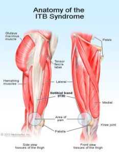
ITBSigns/Symptoms

Runners usually have no exact history of trauma and find that the pain comes on gradually over the outside of the knee during a run. The pain usually appears within a few km of a run and increases in intensity. That same area can also be tender to touch.

Pathophysiology/Etiology

There are a couple theories on the pathophysiology of ITBs.

Some researchers believe that ITB inflammation is a result of excessive friction between the ITB and the boney prominences which occur when the ITB slides over the boney structure and causing inflammation during repetitive movements such as running. Others have argued that rather than the ITB band causing excessive friction, the inflammation is caused by the ITB compressing an area of highly innervated fatty tissue between the IT band and boney prominence

Contributing Factors to Developing ITB Syndrome

The most common factor in developing ITBs is an increase in exercise intensity through mileage, hill training or speed work.

Other reported possible causes which may increase tension in the ITB by altering hip and knee angles include:

  • Downhill running
  • Wearing old shoes
  • Always running on same side of road
  • Leg length discrepancies
  • Excessive pronation of the foot (foot rolling inward)
  • Tight ITB
  • Weakness of glute medius muscle

ITBs and Running Biomechanics

It has been suggested that injuries can manifest as a result of an increase in exercise intensity beyond a threshold level, combined with certain intrinsic factors in athletes.

A recent systematic review looked at biomechanical variables and investigated whether distance runners who suffer from or develop ITBs have different biomechanics than runners who do not develop ITBs.

The evidence shows that it is unlikely that abnormal biomechanics at the foot or shin bone can contribute to increasing tension of the ITB.

The results suggest more is happening at the hip and knee. Runners who eventually develop ITBs have more internal rotation at the knee and greater glut medhip adduction angles during the stance phase of running (when the foot is in contact with the ground) compared to healthy controls.
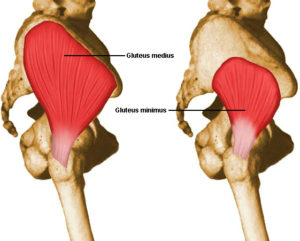
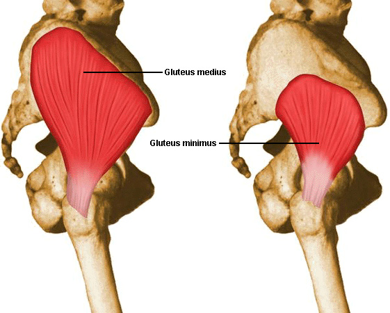
Some researchers have found that this internal rotation of the knee is due more to an externally rotated femur (thigh bone rotated outside) and suggests this may be due to insufficient activity in the medial rotators of the hip (gluteus medius, gluteus minimus, TFL).

As for muscle strength and endurance, there is currently no evidence to suggest that reduced muscle strength plays a role in ITBs. However, the research is limited because many of the trials give inaccurate impressions of a muscle’s functional strength. Future research is needed to look at the timing of muscle action rather than the magnitude of strength.

Research also suggests that runners with current ITBs tend to have more trunk flexion than healthy controls. It is uncertain whether the increased trunk flexion is due to a tight ITB or if the ITB becomes tight as a consequence of the flexed trunk (aka: the torso leaning forward)

How do we treat it!?

Stay tuned to my next article on the latest evidence for treating and managing ITBs!

References:

Foch et la. Associations b/n IT band injury status and running biomechanics in women. Gait & Posture. 2014.

Louw & Deary. The biomechanical variables involved in the aetiology of iliotibial band syndrome in distance runners – a SR. Physical Therapy in Sport. 2014
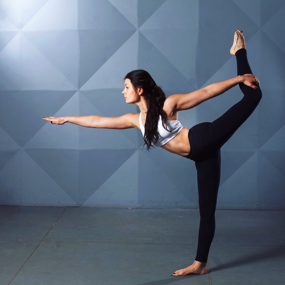
Let’s target the hip abductor muscles! In my last article I discussed the importance of core strength and control to stabilize our trunk when we run – specifically the importance of the hip abductor muscles.

To refresh, the hip abductors help to bring the leg to the outside of our midline to counteract the moment of force where our leg naturally wants to move toward the midline each time our foot hits the ground.

We have several hip abductor muscles in our body, the majority of them are in our butt muscles! Gluteus medius (Gmglut meded), gluteus maximus (GMax), gluteus minimus (GMin) and TFL (tensor fascia latae). The focus of this article is going to be on the gluteus medius because of its important role in stabilizing the pelvis.
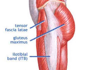
Why do the gluteal muscles become weak? Most of us spend our days sitting and therefore develop weak gluteal muscles making it harder to recruit them during exercise. This can lead to improper use and poor muscle patterning of other muscles to try to compensate for a weak GMed which may increase risk of injury somewhere down the road.

Using techniques to help isolate these muscles can increase their activation and ultimately improve performance. So, how do we target these muscles? A few electromyographic (EMG) studies can help us out!

Study #1: One leg vs. two legs for Gmed activation 

This study used EMG signal amplitude to measure GMed activation in 5 different weight-bearing exercises; double leg stance, single leg (SL) stance, single leg squat, single leg stance on a cushion, and single leg squat on a cushion, where the cushion was an unstable surface underneath the foot to make the exercise more difficult.

To no surprise, the results showed that a SL stance placed higher demands on the GMed than double leg stance and SL squats are more demanding than SL stance. As for the SL exercises on the cushion; the GMed muscle was activated more, but not significantly more than on flat ground.

Study #2: How do we activate Gmed even more?

This study used EMG signals to measure muscle activation patterns of the GMed (among 3 other hip muscles) during 5 unilateral weight-bearing exercises as shown here:
picss

They compared the level of activation to the subject’s maximal voluntary contraction. The EMG signal amplitude had to be between 40-60% of the maximal voluntary contraction to have sufficient intensity for strengthening.

Of the 5 exercises, the results for the GMed showed the highest amount of activation during the wall squat! The next exercises for activation were as follows: forward step-up, lateral step-up, backward step-up and then mini squat. The authors suggested that these exercises may be used as progression exercises towards the wall squat.

TFLStudy #3: What happens when the gluts are weak?

The last article looked at hip abductor muscle activity during resisted side-stepping exercises in either a squatted or standing position. They found that both the GMax and GMed had greater muscle activity during the squatted position than the upright posture. By being in this squatted position, the TFL muscle is less active which means the gluteal muscles should, in theory, be more active. This is important because if the gluteal muscles are weak, the
TFL will compensate which may lead to further underuse and weakening of the gluteal muscles.

Practical Applications

Here are a few key take home points for activating the Gmed to help enhance our running:

  • The GMed plays an important role in stabilizing the pelvis/hip joint during weight-bearing
  • GMed activation is greater when the base of support is less ie: during a side bridge, unilateral squat and lateral step up
  • Some key exercises to get the most activation from the GMed:
    • Single Leg Stance
    • Single Leg Wall Squats
    • Forward Step Ups
    • Lateral Step Ups
    • Side Steps with a resistance band around the knees/ankle (aka: Monster Walks)

And does strengthening GMed actually help to prevent injuries?  As always, the answer is that it depends, but check out this article  from Dr. Delanghe exploring how GMed strength work can decrease injury-causing variability of motion at the knee.

Happy strengthening!

Book your appointment

Contact us to book your next appointment

Call Us 519 885 4930